Agarwal D* and Bansal A
GLA University, Uttar Pradesh, India
- *Corresponding Author:
- Diwakar Agarwal
Department of Pathology
GLA University, Uttar Pradesh, India
E-mail: diwakar.agarwal@gla.ac.in
Accepted date: August 30, 2017
Abstract
Health presents a great challenge for all nations. Improving the health of a nation’s citizens can directly affect the economic growth. Even developed countries, where government spend huge amount of money for establishing medical care units, are found more or less in the custody of various hazardous diseases. Similarly, developing countries particularly like India is facing widespread occurrence of non-communicable disease such as diabetes. Almost 60 million people are diabetic in India. According to the International Diabetes Federation (IDF), diabetes took 0.9 million deaths in 2015 in comparison to other non-communicable diseases. In order to combat the situation, healthy diet plan & physical activity followed by regular check- ups are required to prevent and control diabetes. If not, then the raised blood glucose concentration which is the main cause of diabetes imposes a severe damage to nerves & blood vessels. Among its effects, Diabetic Retinopathy (DR) is the common complication among 10-20% of diabetic subjects. An intervention of Computer Aided Design (CAD) & computer vision technique opens new dimension for medical practitioners in the diagnosis of various abnormalities. The main objective of this literature is to be acquainted with non-invasive methods or procedures for the determination of blood glucose level and screening of DR. Emphasis has been put on mentioning experimental data, applied methodology, clinical applications, and shortcomings.
Keywords
Burden of diabetes, CAD, Glucose, Non-invasive glucose monitoring, Retinopathy
Introduction
Earlier, determining the health status of a person was the prime most challenge before medical experts. In 1940, first blood test was invented for the diagnosis of anaemia. This is one kind of invasive method which is more prevalent nowadays. There are some consequences of it like the subject experiences pain, faces trauma and sense the fear of other type of complications too. Also one has to wait for 2-3 days or 1 week in some cases for the out-coming of diagnosis report. Beside various effects, this method is widely accepted as gold standard method and considered as a reference for validation of several non-invasive diagnostic techniques. However, whether it is the matter of inaccessible complex organs or some common medical conditions like high/low glucose level, blood pressure, jaundice etc almost all medical centers were facing the scarcity of fast, accurate and non-invasive devices.
Non-invasive medical procedure neither requires exploratory surgery nor requires any kind of incision. Numerous impressive non-invasive devices are available in the market. Some of them are already used by physicians generally such as stethoscope (for listening of heart and breath sounds), thermometers (for body temperature examination), sphygmomanometer (for measuring blood pressure) etc. Beside these, many hand held devices like sensor based smart watch and smartphone that are easy to use are also available, so that a person gets health status in a fraction of second.
In recent years, there has been a tremendous research in the field of design and development of Computer Aided Diagnostic (CAD) tools. These tools work on the basis of several imaging modalities such as X-Ray, Magnetic Resonance Imaging (MRI), Computerized Tomography (CT) scan, ultrasound, optical imaging etc. The nightmare of diabetes coerced the inventors and researchers for continuously developing techniques, algorithms, experimental set-ups for the determination of blood glucose level. Through this, severity can be prevented by the timely screening of various disorders such as atherosclerosis, peripheral neuropathy, foot problems, cataract, glaucoma etc caused due to diabetes. Early detection of DR through computer vision has already achieved higher accuracy. This involves pattern recognition, classification and analysis of fundus retinal images.
This literature has been organized into four sections. Section II discusses few commercially available non-invasive devices for the monitoring of blood glucose elevation, blood pressure and bilirubin concentration. Section III focuses on several CAD tools for the determination of blood glucose level. Section IV describes the methods available for the early detection of most occurring effect of diabetes i.e. DR through computer vision.
Non-invasive devices/gadgets for the monitoring of common medical conditions
Several glucose monitoring devices are available in the market. Dexcom G5 Continuous Glucose Monitoring System (CGM) manufactured by Dexcom Inc as shown in Figure 1a and active blood glucose meter manufactured by Accu-Chek as is shown in Figure 1b are in vogue currently. These devices help us to monitor and track glucose level continuously and remotely. Doctors also adjust the therapy for the subject according to the displayed information. Smartphone apps together with pain less non-invasive devices track everything. It calculates how much insulin we need, set reminders, and also prepare graphs and data sheet of our data. One can easily download data on computer, tablet, or smartphone to see patterns and trends in his/her sugar levels.
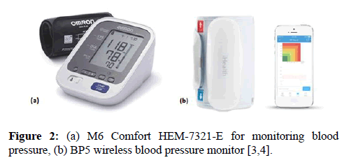
Different types of blood pressure monitoring devices are available for controlling hypertension which is the main risk factor for stroke. Omron’s M6 Comfort HEM-7321-E, which is shown in Figure 2a reduces the inaccuracy caused by incorrect cuff positioning. Other than this, wireless BP5 as shown in Figure 2b manufactured by iHealth gives technology a new wing. It removes all wires and tubes and transfers the data to the smartphone through Bluetooth. The person records the data, share it with friends and doctor on iHealth MyVitals application.
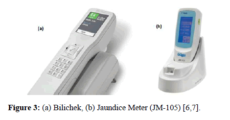
Diagnosis of jaundice in neonates is done by Transcutaneous Bilirubinometer (TcB). Yamanouchi et al. introduced first TcB in the year 1980 together with Minolta Camera Company [5]. TcB emits light on the neonate’s forehead or upper part of sternum and measures the spectral reflectance by the bilirubin component. Currently the two most popular TcBs: Bilichek by Philips Healthcare as shown in Figure 3a and Jaundice Meter JM-102/JM-103/JM-105 by Draeger Medical Inc. are available commercially. JM-105 is shown in Figure 3b.
Non-invasive measurement of blood glucose concentration
Diabetes Mellitus (DM) (also called diabetes) is caused due to abnormal elevation of glucose level and low insulin level in the blood. Badly controlled diabetes leads to extreme complications such as foot ulcers, skin disorders, ischemic heart disease, hypertension, hearing loss, Kidney failure, diabetic retinopathy etc [8]. It is mentioned in World Health Organization (WHO) report that global pervasiveness of diabetes has raised from 4.7%-8.5% since 1980 in adult population [9]. Recently type 2 diabetes has increasingly been reported in children and adolescents [10].
Most of the methods for the determination of blood glucose concentration are invasive. Typically, Glucose oxidase (Gox) method is used. In this method blood samples are taken from the subject. In order to maintain glucose concentration at a desired level, testing is done several times including before and after meal. Although traditional method gives proper result but it has some limitations. It is very painful to take blood several times and also costly due to unaffordable Gox reagents which are used in hospitals. Additionally, sometimes it may contaminate the blood which is the main cause of serious infection [11].
Therefore, researchers continuously work to develop noninvasive methods for the determination of blood glucose concentration. Although non-invasive concept was invented about three decades ago, a large number of techniques are still waiting to launch for the development of health sensing and care. In literatures, description of several non-invasive methods is available. However, the scope is so broad that novel techniques are continuously invented year by year together with the outdating of previous information. In 2012, So et al. provided an overview, advantages and limitations of various non-invasive modalities in view of blood glucose measurement [12]. Emphasis has been put on describing Bioimpedance Spectroscopy, Electromagnetic sensing, Fluorescence technology, Mid-infrared spectroscopy, Near infrared spectroscopy, Optical Coherence Tomography (OCT), optical polarimetry, Raman spectroscopy, Reverse iontophoresis and Ultrasound technology. After that new techniques have been reported in this manuscript.
Ashok et al. presented a new method by using optical monitoring system [13]. The system includes 632.8 nm (Nanometer) wavelength helium neon laser sources of 5 mW power, photo detectors and digital storage oscilloscope. The received signals are then decomposed using Harr wavelet transform. Prediction of glucose level is done using Back Propagation Network (BPN) with gradient descent algorithm and Radial Basis function (RBF). The method was tested on 900 individuals and shows a remarkable difference between DM and non-DM subjects. Efficiency reached up to 99.136% through BPN and 99.53% through RBF. Ahmad at al. used near infrared transmittance spectroscopy across the ear lobe to measure blood glucose level [14]. Transmittance depends upon several physical parameters such as amount of glucose in the blood, amount of blood present in the path of light and thickness of ear lobe tissue. Attenuated lower, middle, and higher wavelength signal is received, sampled and processed using programmable system-on-chip (PSoC-5LP). Oxygen and glucose concentration are then displayed using best fit regression analysis. Total 120 samples were captured and accuracy was reached to 85% when compared to non-invasive glucometer.
Zongyan He invented another method which is based on the metabolic heat measurement [11]. Increased glucose level increases body heat due to the acceleration of metabolic process. The concept was based on the relationship between probe temperature and blood glucose concentration. Correction of several undesirable factors like environmental temperature, humidity, basal body temperature, the pulse and other physiological parameters are taken into account. Pikov et al. found another way based on the transmission of electromagnetic waves of millimetre wavelength range through human skin [15]. Milli-Microwaves (MMW) of 20-300 GHz was used in the experiment. Glucose concentration was calculated by noticing the changes in amplitude and phase of received MMW. Bansal et al. described a new method for determining diabetes through iridology by utilizing iris recognition system [16]. The process was applied on 40 diabetic and 40 non-diabetic subject’s left eye. Segmentation, feature extraction and classification were done by circular Hough transform, 2-d Discrete Wavelet Transform (DWT) and Support Vector Machine (SVM) respectively. Later, efficacy of the process was proved by using certain performance evaluators like sensitivity (95%), specificity (90%) and accuracy (87.5%). Table 1 illustrates the comparison among various methods.
| S. No | Author | Method Used | Accuracy | Experimental Data |
|---|---|---|---|---|
| (%) | ||||
| 1 | Ashok et al. [13] | Optical System | 99.136 | 900 subjects |
| 99.53 | ||||
| 2 | Ahmad et al. [14] | Near Infrared Transmittance Spectroscopy | ||
| 85 | 120 subjects | |||
| 3 | Zongyen He [11] | Metabolic heat measurement | – | – |
| 4 | Pikov et al. [15] | MMW Spectroscopy | 40 DM and 40 Non-DM subjects | |
| 87.5 | ||||
| 5 | Bansal et al. [16] | Iris image | – | – |
Table 1: A comparison of different methods used for measurement of blood glucose concentration.
Non-invasive screening of DR
DR is a disease mainly caused due to retinal disorder or retinal abnormality. DR is the leading ophthalmic pathological cause of blindness among the people of working age in developed countries [17]. Increasing universality of type 2 diabetes give rise to vision threatening DR. It is estimated that 15 to 25% of the diabetic population have DR [18]. Usually, DR subjects do not show any type of symptoms at an early stage until vision loss at later stages. Therefore, immediate actions by the subject need to be taken if he/she encountered high blood glucose level in their first time check-up. The early stage of DR i.e. Non- Proliferative Diabetic Retinopathy (NPDR) develops leakage of extra fluid and small amount of blood into the eye. This leads to appear Microaneurysm, hemorrhages, hard exudates, macular edema, and macular ischemia. The later stage of DR i.e. Proliferative Diabetic Retinopathy (PDR) mainly occurs due to the closure of blood vessels. This gives birth to new vessels for blood supply called neovascularization [19].
Salz et al. provided a summarized description and limitations of various imaging modalities used for the treatment and diagnosis of different forms of DR [20]. Fluorescein Angiography (FA), B-scan ultrasonography, OCT and digital color fundus photography were described significantly. FA can show microaneurysms in retina, Intra-Retinal Microvascular Abnormalities (IRMA), neovascularization and macular edema. The potential side effects are allergy, nausea and sometimes vomiting. B-scan ultrasonography assists in the diagnosis of PDR. It can also show retinal detachment and vitreous hemorrhage. B-scan ultrasonography is not helpful if media opacity is clear. OCT is capable of capturing high-resolution images and can determine retinal thickness and loss of different layers of retina. It is helpful in the management of macular edema. OCT does not assist in the diagnosis of macular ischemia.
There has been a huge amount of research in the diagnosis of DR through computer vision. One of the main reasons behind its usefulness is a transparency between subject and medical practioner about the treatment. Fundus photography demonstrates the subject about the nature and form of disease. With the help of fundus images, physicians regularly monitor the progressiveness of DR over time. Three types of fundus photography are mainly encountered: standard, wide-field and stereoscopic [20].
DM has been referred as a vascular disease because it affects vascular endothelial wall. Since many years, analysis of blood vessels plays a very crucial role in the screening and diagnosis of various eye diseases. Assessment of the characteristics like vessel width, tortuosity, abnormal branching etc helps in understanding cataract, glaucoma, DR and other diseases more precisely. Moreover, determination of proper location of blood vessels may help medical practitioners in finding correct other fundus features such as optic disk and fovea. Besides these analysis, premature retinopathy can be screened through vascular tree segmentation and narrowing of arterioles [21–23], degree of hypertensive retinopathy can be determined through the characterization of vessels tortuosity [24], hypertension and cardiovascular diseases can be diagnosed through vessel diameter measurement [25–27] and many more. People with both type 1 and type 2 diabetes are at risk of developing DR. Diabetic Retinopathy may not have any symptoms or may not affect sight in the early stages [28]. Therefore, if the condition is caught early, then the treatment is more effective for reducing or preventing the damage to sight. Regular screening for DR is important if anyone have diabetes. In view of this, several methods are available for the segmentation of retinal blood vessels [29–32]. Emphasis has been put on automatic depiction and analysis of the characteristics of blood vessels.
Bansal et al. determines retinal blood vessels width and orientation for the diagnoses of glaucoma [30]. Hassan et al. proposed a method for the detection of neovascularization [32]. New blood vessels are grown due to excessive lack of oxygen in retinal capillaries. Neovascularization is the worst case of retinal abnormality. Detection of it gives a new path in medical science for better understanding the retinal disorders.
Although assessment of blood vessels hold an important role in diagnosing vascular diseases, detection and understanding of lesions like microaneurysm, exudates and hemorrhages is needed for the screening of DR. Sopharak et al. develops an automatic screening system for DR by detecting and segmenting microaneurysm in digital fundus photographs [33]. Mathematical morphology was taken into account and performance was measured on 45 non-dilated retinal images. Sensitivity (81.61%), specificity (99.99%), precision (63.76%) and accuracy (99.98%) were determined for efficacy of the method. Later, they classify degree of DR from mild to severe on the basis of number of microaneurysm reported. Akram et al. proposed an identification and classification of microaneurysm for the early detection of DR [34]. Gabor filter was used for the enhancement of microaneurysm to make it distinguishable from other type of lesions. Various feature vectors and a hybrid classifier, which is a combination of Guassian Mixture Model (GMM) and SVM were formed on the basis of color, shape and size. Sensitivity (98.64%), specificity (99.69%) and accuracy (99.40%) were calculated for 219 retinal images for the purpose of evaluation.
Mahendran et al. detects and segment the exudates from low contrast retinal images. Further investigation on the severity level of DR was done by using SVM and Probabilistic Neural Network (PNN) classifiers [35]. GLCM based features of 370 retinal images were extracted and on the basis of that, SVM attained the sensitivity, specificity and accuracy of 98.68%, 100% and 97.89% respectively. In contrast PNN achieved sensitivity, specificity and accuracy of 96.64%, 98.46% and 94.76% respectively. Romero et al. applied Principal Component Analysis (PCA) for the detection of microaneurysm in order to early diagnosis of DR [36]. Two dataset of 89 and 100 retinal images were taken. First dataset reported sensitivity (92.32%), specificity (93.87%) and accuracy (95.93%). In contrast second dataset reported sensitivity (88.06%), specificity (97.47%) and accuracy (92.19%). Haloi et al. proposed a novel method based on Deep Neural Network (DNN) [37]. Here, three data sets of 50, 1200, and 89 retinal images were formed. Each pixel is classified as microaneurysm or non-microaneurysm without need of preprocessing and feature extraction procedures. Sensitivity (87.0%), specificity (95.0%) and accuracy (98.8%) were achieved. Table 2 illustrates a comparison among various methods.
| S.No | Author | Method/Classifier Used | Sensitivity | Specificity | Accuracy | Experimental data |
|---|---|---|---|---|---|---|
| (%) | (%) | (%) | ||||
| 1 | Sopharak et al. [33] | Mathematical Morphology | 81.61 | 99.99 | 99.98 | 45 non-dilated images |
| 2 | Akram et al. [34] | GMM and SVM | 98.64 | 99.69 | 99.4 | 219 retinal images |
| 3 | Mahendran et al. [35] | SVM | 98.68 | 100 | 97.89 | 370 retinal images |
| PNN | 96.64 | 98.46 | 94.76 | |||
| 4 | Romero et al. [36] | PCA | 92.32 | 93.87 | 95.93 | 89 retinal images |
| 88.06 | 97.47 | 92.19 | 100 retinal images | |||
| 5 | Haloi et al. [37] | DNN | 87 | 95 | 98.8 | Three data sets of 50, 1200, and 89 retinal mages. |
Table 2: Comparison chart of non-invasive methods for the screening or early detection of DR.
Not only DR but diseases related to stomach problems, foot problems, high blood pressure, stroke and bacterial infections are also occur due to diabetes. Early detection of such diseases can prevent the severe complications in diabetic subjects. Computer Aided Diagnostic (CAD) systems need to be developed. The increasing cost of medicine and insufficient resources for development boost up the necessity for such an approach.
Conclusions
Several methods used for the non-invasive measurement of glucose concentration have been studied. Insight of these methods opens new dimension for thinking about other non-invasive procedures. Moreover, there has been a constant demand for techniques which are based on precise mathematical modeling. In view of vast applications of color image processing, one can utilize iridology and sclerology for the better understanding of diabetes and its health hazardous effects. Analysis of color components of palpebral conjunctiva can also paved a new path in the determination of blood glucose concentration. Efficiency of existing method needs to be improved in order to get accurate information. So that medical practitioner makes the best plan for managing the diabetes.
Acknowledgement
Diwakar Agarwal researched the data and wrote the manuscript. Atul Bansal reviewed the manuscript and give suitable suggestions.
Diwakar Agarwal is the guarantor of this work and, as such, had full access to all the data in the study and takes responsibility for the integrity of the data and the accuracy of the data analysis.
References
- Dexcom G5 CGM System. Dexcom Inc.
- Accu-Chek Active Blood Glucose Meter. Accu-Chek.
- M6 Comfort HEM-7321-E. Omron.
- BP5 wireless blood pressure monitor. iHealth.
- Yamanouchi I, Yamauchi Y, Igarashi I, et al. Transcutaneous Bilirubinometery: Preliminary Studies of Non-invasive Transcutaneous Bilirubin meter in the Okayama National Hospital. Pediatrics. 1980;65:195-202.
- Bilichek. Philips Healthcare.
- Jaundice Meter (JM-105). 2017.
- Diabetes: Symptoms, Causes and Treatments. 2016.
- Global Report on Diabetes. Geneva: World Health Organization. 2016.
- Beat diabetes: Scale up prevention, strengthen care and enhance surveillance. India: World Health Organization. 2016.
- He, Z. Method for Non-invasive Blood Glucose Monitoring. US. Patent 8,676,284 B2, New Jersey: US, 18 March 2014.
- So CF, Choi KS, Wong TK, et al. Recent Advances in Non-invasive Glucose Monitoring. Medical Devices (Auckland, NZ). 2012;5:45-52.
- Ashok V, Kumar N. Determination of Blood Glucose Concentration by Using Wavelet Transform and Neural Networks. Iran J Med Sci. 2013;38:51-6.
- Ahmad M, Kamboh A, Khan A. Non-invasive Blood Glucose Monitoring Using Near- Infrared Spectroscopy. 2013.
- Pikov V, Siegel PH. Methods and Systems for Non-invasive Measurement of Blood Glucose Concentration by Transmission of Millimeter Waves through Human Skin. Patent 20160051171A1, California: US. 2016.
- Bansal A, Agarwal R, Sharma RK. Determining Diabetes Using Iris Recognition System. Int J Diabetes Dev Ctries. 2015;35:432-8.
- Taylor HZ, Keeffe JE. World blindness: A 21st century perspective. Br J Ophthalmol. 2001;85:261-66.
- Guidelines for the comprehensive Management of Diabetic Retinopathy in India. A vision 2020 The Right to Site India Publication. 2008.
- Diabetic Retinopathy. 2016.
- Salz DA, Witkin AJ. Imaging in Diabetic Retinopathy. Middle East Afr J Ophthalmol. 2015;22:145-50.
- Heneghan C, Flynn J, O’Keefe M, et al. Characterization of Changes in Blood Vessel Width and Tortuosity in Retinopathy of Prematurity Using Image Analysis. Med Imag Anal. 2002;6:407-29.
- Grisan E, Ruggeri A. A Divide and Impera Strategy for the Automatic Classification of Retinal Vessels into Arteries and Veins. In: Proceedings of the 25th international Conference IEEE Eng. Med Biol Soc. 2003;890-93.
- Hatanaka Y, Fujita H, Aoyama M, et al. Automated Analysis of the Distribuitions and Geometries of Blood Vessels on Retinal Fundus Images. In: Proceedings of the SPIE Med Imag Process. 2004;5370:1621-8.
- Foracchia M, Grisan E, Ruggeri A. Extraction and Quantitative Description of Vessel Features in Hypertensive Retinopathy Fundus Images. In: Book Abstracts of the 2nd International Workshop Comput. Asst. Fundus Image Anal. 2001; 6.
- Goa X, Bharath A, Stanton A, et al. A Method of Vessel Tracking for Vessel Diameter Measurement on Retinal Images. In: Proceedings of ICIP. 2001;881–4.
- Martinez Perez ME, Hughes AD, Stanton AV, et al. Retinal Vascular Tree Morphology: A Semiautomatic Quantification. In: IEEE Transaction of Biomed. Eng. 2002;49:912-7.
- Lowell J, Hunter A, Steel D. Measurement of Retinal Vessel Widths from Fundus Images Based On 2-D Modeling. In: IEEE Transaction of Med. Imag. 2004;23:1196-1204.
- Diabetic Retina Screen-The National Diabetic Retinal Screening Programme. 2016.
- Marín D, Aquino A, Bravo JM, et al. A New Supervised Method for Blood Vessel Segmentation in Retinal Images by Using Gray-Level and Moment Invariants-Based Features. In: IEEE Transactions on Medical Imaging. 2011;30:146-57.
- Bansal N, Kuthirummal S, Eledath J, et al. Automatic Blood Vessel Localization in Small Field of View Eye Images. In: Proceedings of the 32nd Annual International Conference of the IEEE EMBS. 2010;5644-8.
- Salazar Gonzalez A, Kaba D, Li Y, et al. Segmentation of the Blood Vessels and Optic Disk in Retinal Images. IEEE Journal of Biomedical and Health Informatics. 2014;18:1874-86.
- Hassan SSA, Bong DBL, Premsenthil M. Detection of Neovascularization in Diabetic Retinopathy. J Digit Imaging. 2012;25:437-44.
- Sopharak A, Uyyanonvara B, Barman S. Automatic Microaneurysm Detection from Non-dilated Diabetic Retinopathy Retinal Images Using Mathematical Morphology Methods. IAENG Int J Comput Sci. 2011;2:38.
- Akram MU, Khalid S, Khan SA. Identification and Classification of Microaneurysms for Early Detection of Diabetic Retinopathy. J Pattern Recognition. 2013;46:107-16.
- Mahendran G, Dhanasekaran R. Investigation of the Severity Level of Diabetic Retinopathy Using Supervised Classifier Algorithms. J Comput Electr Eng. 2015;45:312-23.
- Romero RR, Harnandez J. A Method to Assist in the Diagnosis of Early Diabetic Retinopathy: Image Processing Applied to Detection of Microaneurysms in Fundus Images. Comput Med Imaging Graph. 2015;44:41-53.
- Haloi M. Improved Microaneurysm Detection Using Deep Neural Networks. J Computer vision and pattern recognition. 2016.


Leave A Comment